Reconstruction of transparent objects using phase shifting profilometry based on diffusion models
Reconstructing the 3D surface of transparent objects with phase shifting profilometry is difficult because refraction and internal reflections corrupt the sinusoidal fringe patterns at and near the object surface. Existing phase unwrapping and depth recovery algorithms assume clean fringe data and produce large errors or missing regions when the input is corrupted by transparency. This paper proposes using denoising diffusion probabilistic models (DDPMs) as a learned prior over plausible phase maps, conditioned on the observed corrupted fringe images. The iterative denoising process of the diffusion model naturally handles the inpainting and denoising sub-problems jointly, guided by the corrupted observation at each step. The method is evaluated on a diverse set of transparent objects including glass components and plastic parts common in industrial inspection. Results published in Optics Express (2024) show substantial improvement in reconstruction completeness and surface accuracy compared to classical phase retrieval and prior CNN-based approaches, establishing diffusion models as a promising tool for challenging optical measurement scenarios.
Problem setting
Transparent objects present a well-known challenge for phase shifting profilometry: refractions and specular reflections corrupt the captured fringe patterns in ways that violate the assumptions of standard phase retrieval algorithms, leading to missing or inaccurate depth regions. This work proposes using diffusion models—a class of generative models with state-of-the-art image synthesis capability—to reconstruct transparent object surfaces from corrupted profilometry data. The diffusion model is conditioned on the corrupted fringe observations and learns to iteratively denoise and inpaint phase maps to produce physically plausible surface reconstructions.
The figures below collect representative visual evidence from Optics Express, 32(8):13342–13356.
Method and visual evidence
The figures below summarize the paper’s setup, signal flow, and visual evidence.
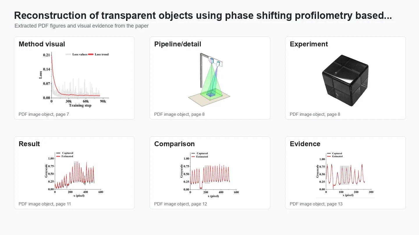
Method overview.
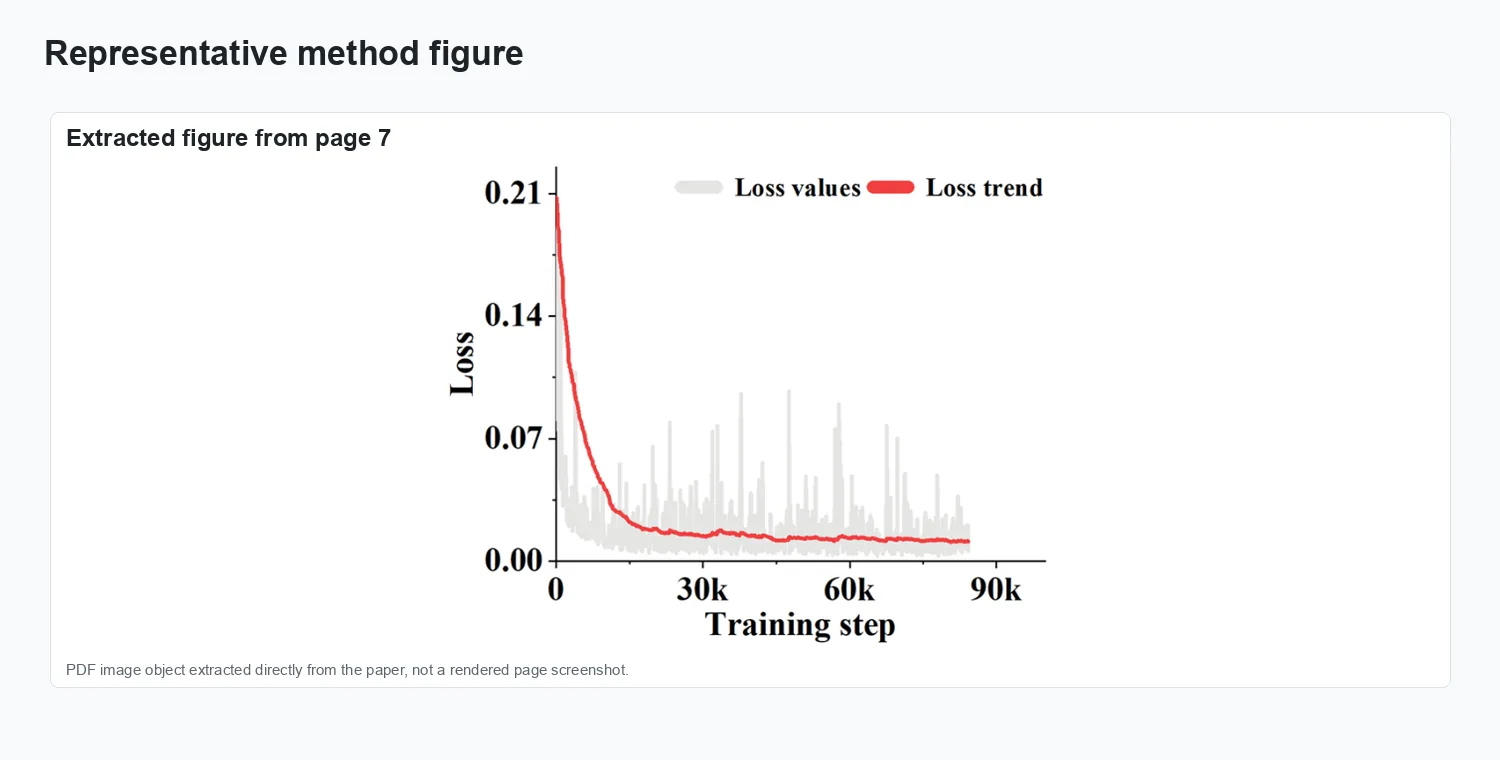
Representation and setup.
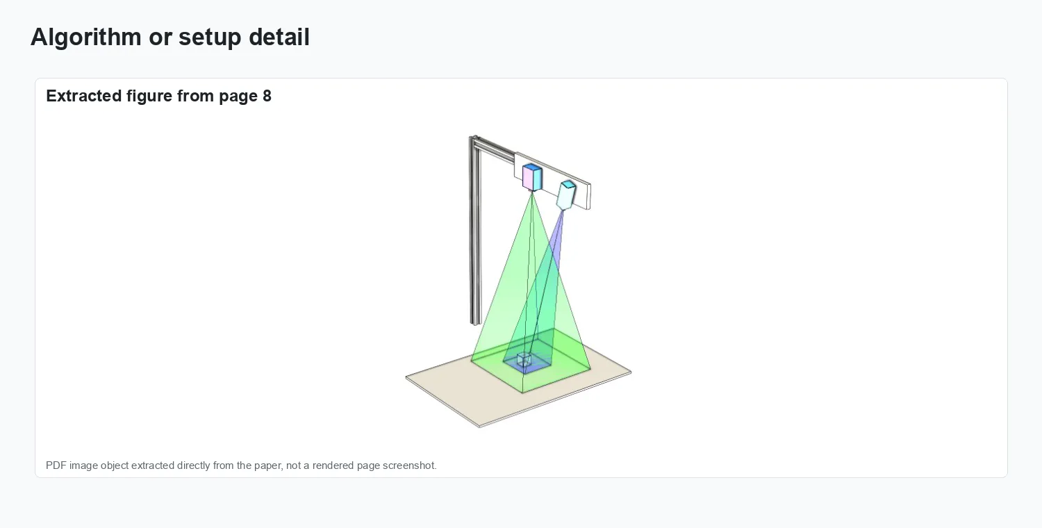
Experimental evidence.
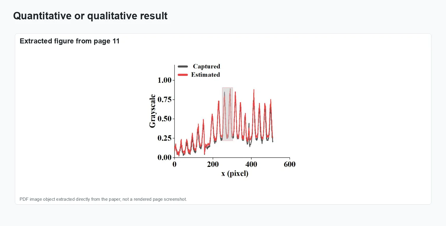
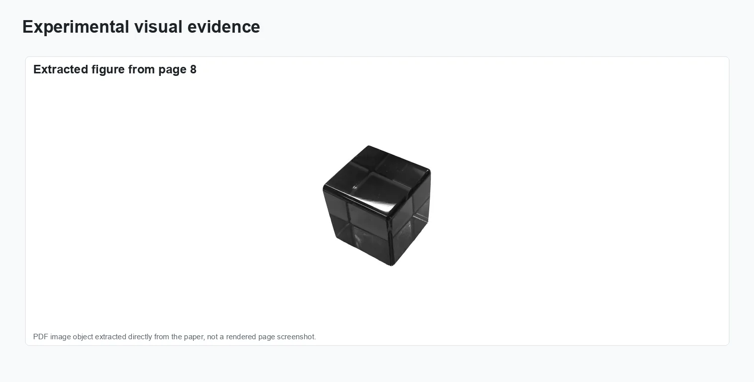
Result comparison.
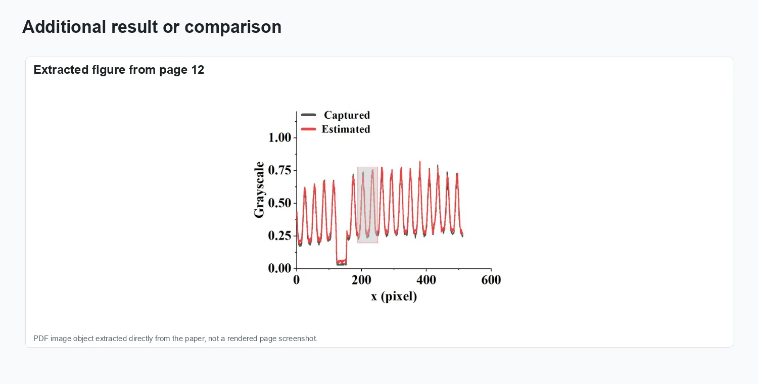
Additional visual result.
Results and impact
The evaluation reported in Optics Express, 32(8):13342–13356 is summarized through the figures above.